Nano-Structure Physiology
RESEARCH THEMES
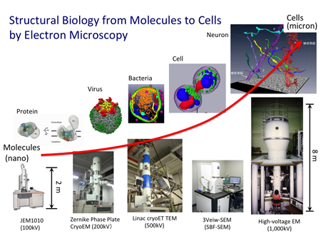
Our research goal is to reveal the relationship between biological functions and structures. For this purpose, we use different types of state-of-the-art electron microscopes (Figure). First, the Zernike phase-plate electron cryomicropscope with energy filter (JEM-2200FS: 200kV) is used for high-resolution structural analyses of non-stained biological samples. Second, the high voltage electron microscope (HVEM) for biologcal research (H-1250M: 1MV) equipped with a direct detection CMOS camera is employed to visualize the three-dimensional structures of thick biological specimens, like cell and neuron. The serial block-fase SEM is also used for the further thick materials, like tissues and neural circuits. By using these microscopes, we carry out a structural biology from molecules to cells.
SELECTED PUBLICATIONS
- "Structural basis for broad detection of genogroup II noroviruses by a monoclonal antibody that binds to a site occluded in the viral particle" J. Virol. 86, 3635-3646 (2012)
- "Three-dimensional structure of the alpha1-beta complex in the skeletal muscle dihydropyridine receptor by single-particle electron microscopy" J. Electron Microsc. 59, 215-226 (2010)
- "Zernike phase contrast cryo-electron microscopy and tomography for structure determination at nanometer and subnanometer resolutions" Structure 18, 903-912 (2010)
- "Structural changes in a marine podovirus associated with release of its genome into Prochlorococcus" Nat. Struct. Mol. Biol. 17, 830-836 (2010).